Vision & Mission
Vision
-
To impart high-quality patient care, academics, and research in the field of Radiology and Imaging to match concurrent global standards.
Mission
-
To offer subspecialty practice for better patient care.
-
To create a center of excellence in Fetal Imaging, MSK Imaging, Neuroradiology, Cross-sectional Imaging, and Interventional Radiology and offer outstanding services to deserving patients and academics to healthcare personnel.
-
To focus on clinical, interdisciplinary, and translational research with the incorporation of technology and artificial intelligence.
Vision
- To impart high-quality patient care, academics, and research in the field of Radiology and Imaging to match concurrent global standards.
Mission
- To offer subspecialty practice for better patient care.
- To create a center of excellence in Fetal Imaging, MSK Imaging, Neuroradiology, Cross-sectional Imaging, and Interventional Radiology and offer outstanding services to deserving patients and academics to healthcare personnel.
- To focus on clinical, interdisciplinary, and translational research with the incorporation of technology and artificial intelligence.
HOD Profile
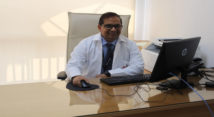
Name:- Dr. Amol Gautam
Qualification:- MBBS, DMRD, DMRE, DNB (Radiodiagnosis),
MNAMS, FRCR(London), EDiR (Spain), DICR
Name:- Dr. Amol Gautam
Qualification:- MBBS, DMRD, DMRE, DNB (Radiodiagnosis),
MNAMS, FRCR(London), EDiR (Spain), DICR
Dr. Amol Gautam has obtained his MBBS degree from prestigious and one of the oldest medical school of India, Grant Medical College and Sir JJ Hospital, Mumbai and his PG diplomas and degrees in the subject of Radiodiagnosis from the same institute. Presently he is serving as a Professor and Head of the Department of Radiodiagnosis at Symbiosis Medical college for Women at Pune.
Experience:
He has over 16 years of experience in patient care (radiology reporting), academics and research activities.
Achievements:
Fellow of Royal College of Radiologists (FRCR) after passing qualifying examination in May 2009. Also, recently passed European Diploma in Radiology (EDiR) examination in April 2022 to update contemporary knowledge. He has served as an examiner in several universities for Post Graduate MD (Radiodiagnosis) examination since 2018. He has guided several post graduate students in their research work and dissertations.
His field of interests are Neuroradiology, Musculo-Skeletal Radiology and Body MR imaging and delivered invited faculty talks in many conferences.
He is serving as an assessor for NABH-Medical Imaging Services (MIS) by Quality Council of India since 2020.
Paper Publications::
He has published more than 19 papers in various National and International journals
Infrastructure facilities
Equipments
|
RADIOLOGY DEPARTMENT |
||||
|
MACHINE NAME |
COMPANY NAME |
DESCRIPTION |
QUANTITY |
|
| X-Ray with Fluoroscopy (DR) | SHIMADZU Corporation, Japan | 800 mA | 1 | |
| X-Ray (CR) | M/s. Allengers Medical Systems Limited, India | 500 mA | 2 | |
| X-Ray (CR) | BPL Medical Technologies Pvt Ltd, India | 400 mA | 1 | |
| X-Ray Mobile (DR) | SHIMADZU Corporation, Japan | 400 mA | 1 | |
| X-Ray Mobile (CR) | BPL Medical Technologies Pvt Ltd, India | 70 mA | 1 | |
| X-Ray Mobile (CR) | BPL Medical Technologies Pvt Ltd, India | 100 mA | 2 | |
| USG & Colour Doppler | GE | Voluson E6, Logiq E10, Voluson S8 | 3 | |
| USG Mobile | GE | Logiq | 1 | |
| CT Scan | PHILIPS | 128 Slice Ingenuity | 1 | |
| MRI | PHILIPS | 3 Tesla Ingenia | 1 | |
| Mammography | HOLOGIC | Digital Mammography with Mammotomosynthesis | 1 | |
| DEXA | HOLOGIC | BMD | 1 | |
| Ortho Pantomograph (OPG) | M/s Trophy | 8 mA | 1 | |
| CR | FUJIFILM | Reader with printer | 2 | |
| DR | FUJIFILM | Retrofit mobile X-ray panel | 1 | |
| PACS | Medsynapse | Barco monitors with high-end workstations for reporting | 10 | |
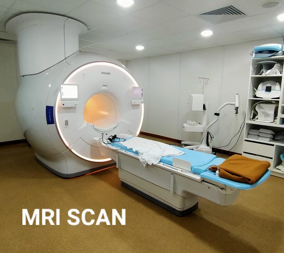
3T MRI
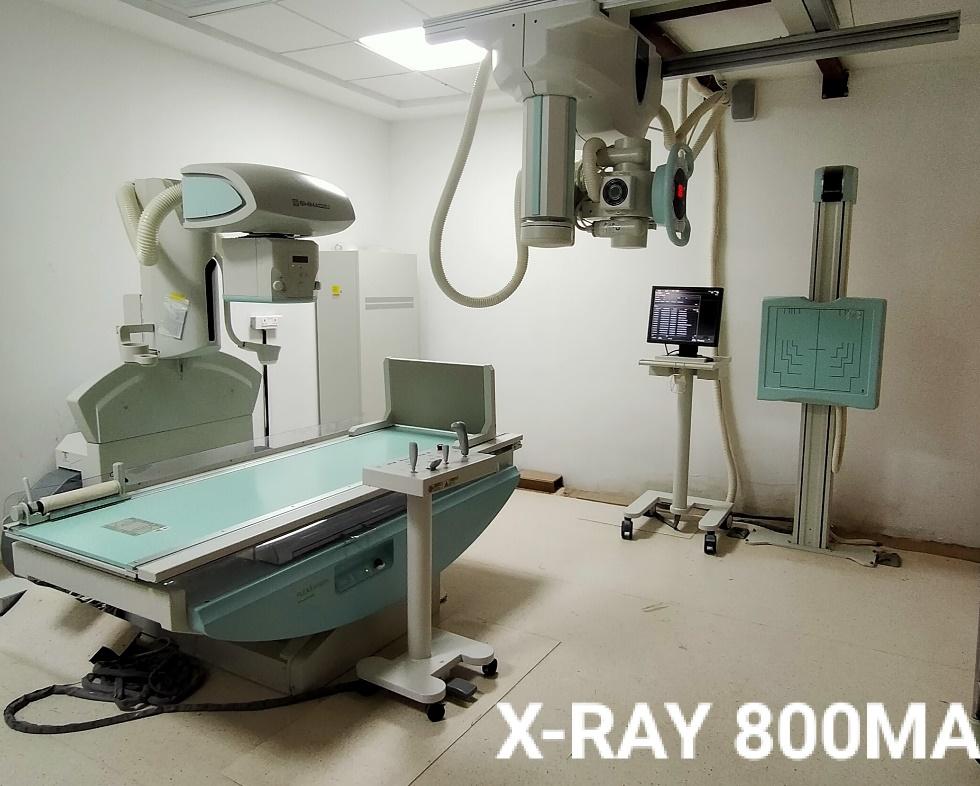
800 mA X-Ray Digital Fluoroscopy
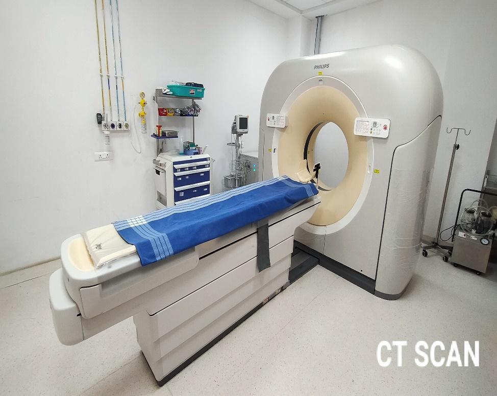
CT Scan – 128 Slice
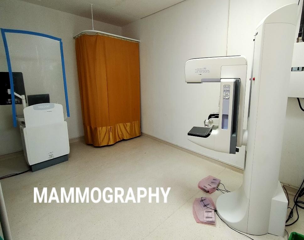
Digital Mammography
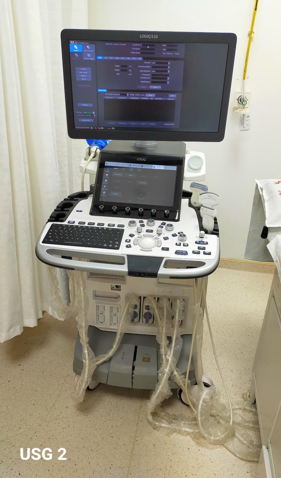
Ultrasonography
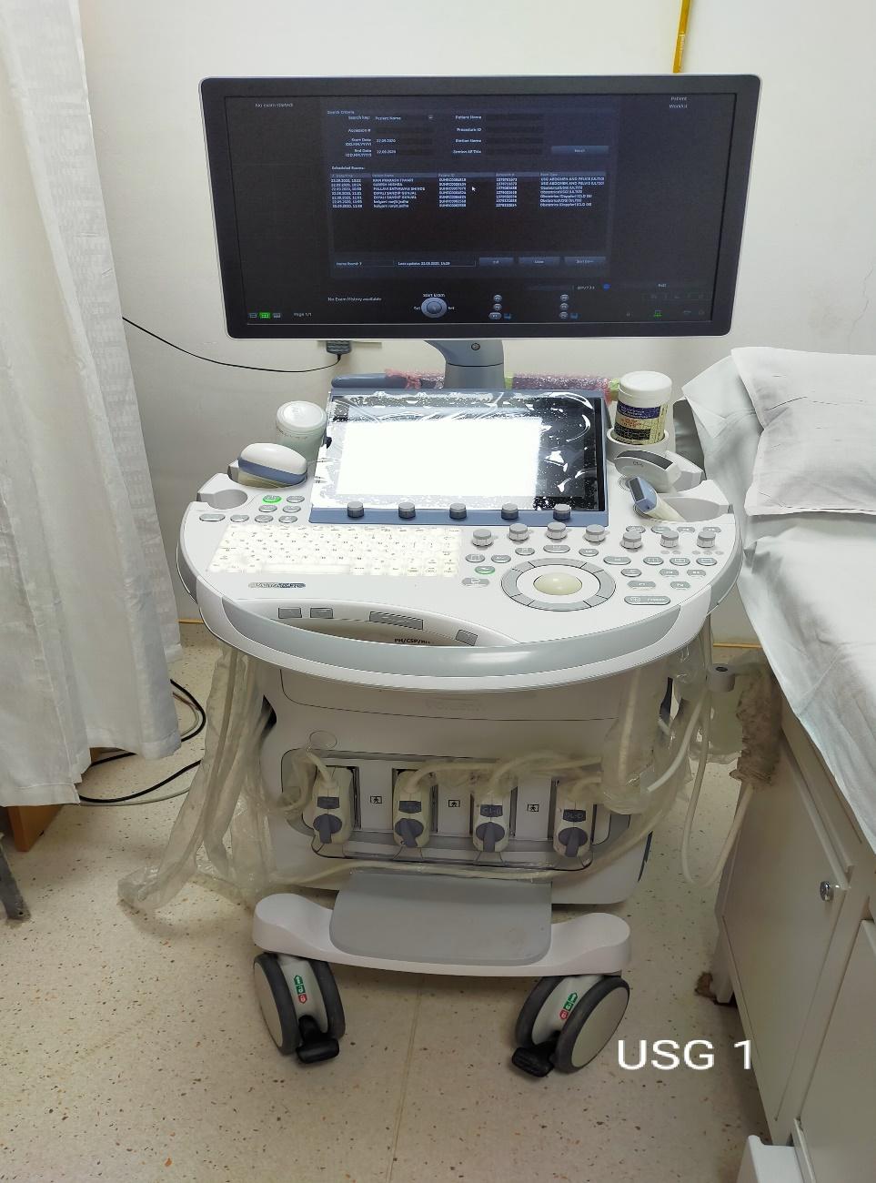
Ultrasonography
Services
Services provided:
- X-Ray - Digital and Computed Radiography, portable radiograph in the ICU and wards
- Screening - Fluoroscopy with guided procedures.
- Special Radiological procedures - Barium studies for bowel and Iodinated contrast studies including HSG, IVP & MCU/RGU Scanogram, etc.
- USG - 3D, volume probes and elastography, Obstetrics USG, Doppler studies
- CT Scan - 128 Multislice CT with dedicated software for cardiac CT coronary angiography, and virtual colonoscopy.
- MRI – 3 Tesla with advanced software e.g. perfusion, spectroscopy, tractography, non-contrast perfusion, T1 mapping, cartography, cardiac MRI with quantification etc.
- Mammography - Digital Mammography with Mammotomosynthesis
- DEXA - Bone Densitometry and Body Fat estimation
- OPG - Dental X-rays
- USG and CT-guided interventions like FNAC, Biopsy, drainage, pig-tail catheter insertion etc.
Research Publications
1. Dr. Amol Gautam
S.N
Year
Original Articles
1
2023
1. Shah SR, Gautam AA, Tamboli AI, Bhoite AS. When formation of cerebral vasculature goes aberrant–A pictorial essay. Medical Journal of Dr. DY Patil University. 2023 Mar 22.
2
2023
2. Kolluru AR, Gautam AA, Garg S, Tamboli AI. Magnetic resonance imaging of synovial diseases of knee. Journal of Datta Meghe Institute of Medical Sciences University. 2023 Apr 1;18(2):291-8.
3
2021
3. Nilesh K, Shah S, Gautam A, Thorat S. Uncontrolled bleeding during tooth extraction from an undiagnosed arteriovenous malformation. BMJ Case Rep. 2021;14(8): e236983.
4
2020
4. Nilesh K, Punde P, Patil NS, Gautam A. Central ossifying fibroma of mandible. BMJ Case Rep. 2020;13(12): e239286.
5
2020
5. Bahekar A, George V, Jacob K, Salih SR, Joseph AT, Koshy C, et al. V-FAT study - A correlation between novel markers of obesity and coronary artery disease severity assessed by Syntax score in patients presenting with acute coronary syndrome. Indian Heart J. 2020;72(5):448–50.
6
2020
6. Gautam A, Wadhwa N. Evaluation of Azygoesophageal Recess in adults with the help of computer based tomography. J. Cardiovasc Disease Res.2020; (2): 159-162.
7
2020
7. Gautam A. Role of MRI in non-traumatic adult chronic hip joint pain. J. Cardiovasc Disease Res. 2020; 11(2): 163-166.
8
2019
8. Hiremath SB, Gautam AA, George PJ, Thomas A, Thomas R, Benjamin G. Hyperglycemia-induced seizures - Understanding the clinico- radiological association. Indian J Radiol Imaging. 2019;29(4):343–9.
9
2019
9. Hiremath SB, Gautam AA, Sasindran V, Therakathu J, Benjamin G. Cerebrospinal fluid rhinorrhea and otorrhea: A multimodality imaging approach. Diagn Interv Imaging. 2019;100(1):3–15.
10
2018
10. Hiremath SB, Gautam AA, Sheeja K, Benjamin G. Assessment of variations in sphenoid sinus pneumatization in Indian population: A multidetector computed tomography study. Indian J Radiol Imaging. 2018;28(3):273–9.
Dr. Uday Zende
1
2024
1.Gupta N, Angadi K, Shukla U, Pathak K, Zende U, Bhakare M. A case report of pulmonary nocardiosis caused by Nocardia otitidiscaviarum. J Krishna Inst Med Sci Univ 2024; 13(1):156-159.
2
2024
2.Datar ML, Jagdale SY, Zende UM. Study of Ultra-Sonographic Evaluation of Vaginal Bleeding in Pregnancy of Maharashtra Population. International Journal of Pharmaceutical and Clinical Research 2024; 16(4); 1122-1126
Dr. Snehil Kumar
1
2022
1.Kumar S, Tamboli AI, Bhoite AS, Bawage RR, Apurva J, Gautam AA.RADIOLOGICAL SPECTRUM OF SINONASAL MUCORMYCOSIS IN COVID POSITIVE PATIENTS. NeuroQuantology. 2022:7365-85.
2
2022
2.Snehil, K., Bhoite, A. S., Asif, T., Bawage, R. R., Garg, S. R., Gautam, A. A., & Shaha, P. R. (2021). COVID-19: HRCT lung pattern, distribution and severity score with clinico-pathological correlation in a tertiary level institution in rural Maharashtra. International Journal of Advances in Medicine, 8(12), 1821.https://doi.org/10.18203/2349-3933.ijam20214517
3
2020
3.Kumar DS, Department of Radiodiagnosis, Krishna Institute of Medical Sciences, Karad, Maharashtra, India. Evaluation of the role of MRCP and ultrasound in diagnosing obstructive jaundice. J Med Sci Clin Res [Internet]. 2020;08(02).
4
2019
4. Adke S, Potdar D, Shaha P, Patil P, Bhoite A, Kumar S, Pansare B. Role of computed tomography in evaluation of renal masses. Journal of Medical Science and Clinical Research. 2019;7(4):791-800.
5
2021
5.Snehil, K., Bhoite, A. S., Asif, T., Bawage, R. R., Garg, S. R., Gautam, A. A., & Shaha, P. R. (2021). COVID-19: HRCT lung pattern, distribution and severity score with clinico-pathological correlation in a tertiary level institution in rural Maharashtra. International Journal of Advances in Medicine, 8(12), 1821.https://doi.org/10.18203/2349-3933.ijam20214517.
Dr. Sharavari Gulve
1
2019
1.Gulve SS, Phatak SV. Parathyroid adenoma: Ultrasonography, Doppler, and elastography imaging.Journal of Datta Meghe Institute of Medical Sciences University. 2019 Jan 1;14(1):47.
2
2020
2.Gulve S, Phatak S, Lohchab B, Patwa P, Tapadia S. Postcesarean section keloid: Ultrasonography,color doppler, and elastography evaluation. Journal of Datta Meghe Institute of Medical SciencesUniversity. 2020 Jul 1;15(3):477.
3
2020
3.Gulve SS, Parihar PS, Dhande R. Role Of Computed Tomography Scan In Evaluation Of Pancreatic Lesions. European Journal of Molecular & Clinical Medicine. 2021 Mar 15;7(11):2020.
4
2021
4.Parihar PH, Gulve SS. Mediastinal Extension of Pancreatic Pseudocyst--A Case Report. Journal of Evolution of Medical and Dental Sciences. 2021 Feb 1;10(5):316-9.
5
2020
5.Gulve SS, Phatak SV, Varma AD, Gupta RS. Importance of Ultrasonography in Inspissated Bile Syndrome in a Neonate--A Case Report. Journal of Evolution of Medical and Dental Sciences. 2020 Oct 5;9(40):3016-9.
6
2021
6.Parihar PH, Gulve SS. Mediastinal Extension of Pancreatic Pseudocyst--A Case Report. Journal of Evolution of Medical and Dental Sciences. 2021 Feb 1;10(5):316-9.
7
2020
7.Patwa P, Phatak S, Lohchab B, Gulve S. Ultrasonography, doppler, and elastograhic imaging of sebaceous cyst of breast. Journal of Datta Meghe Institute of Medical Sciences University. 2020 Jul 1;15(3):474.
8
2020
8.Varma AD, Phatak SV, Gupta RS, Gulve S, Jain S, Sawangi W. Fat Necrosis in Axillary Lipoma after FNAC-A Case Report. Journal of Evolution of Medical and Dental Sciences. 2020 Oct 12;9(41):3070-3.
9
2020
9.Lohchab B, Phatak S, Gulve S, Tapadia S. Intratesticular and extratesticular varicocele: Ultrasound and doppler appearance. Journal of Datta Meghe Institute of Medical Sciences University. 2020 Oct 1;15(4):669-71.
1. Dr. Abhijit Patil
S.N
Year
Original Articles
1.
2021
Patil A M, Meenakshi B, Salvi S. A Novel Severity Scoring derived from Chest Radiographic findings in COVID 19 Patients admitted to a COVID Care University Hospital in India. Journal of Pharmaceutical Research International, 33(37A), 17-25.
2.
2018
Bhargava R, Patil AM, Bakshi V, Kalekar TM, Gandage SG. Utility of contrast-enhanced fluid-attenuated inversion recovery in magnetic resonance imaging of intracranial lesions. West Afr J Radiol 2018; 25:34-8. doi:10.4103/wajr.wajr_4_17
3.
2016
Patil AM, Kalekar TM, Patil PV, Gandage SG. Carotid Doppler in Patients of Stroke. Global J. Res. Anal.2016;5(9):347-351
4.
2016
Kalekar TM , Patil AM. MRI Evaluation of Rotator Cuff and Labral Injuries. Int. J. Sci. Res.2016;5(6):156-158
5.
2016
Kuber R, Randhawa S, Khaladkar S, Patil A, Veeramachaneni R. Doppler study of middle cerebral artery and umbilical artery in biometrically suspected intra uterine growth restricted pregnancies. Int..J. Res. Med. Sci.2016;4(2):403-414.
6.
2016
Patil AM, Kalekar T M, Gandage SG. Computed Tomography Findings in Clinically diagnosed Patients of Stroke.2016;5(5):247-251
7.
2015
Bhatwal AS, Patil AM, Kelkar AB et al. Role of oral gadolinium as a negative contrast medium in Magnetic Resonance Cholangiopancreatography. Int J Health Sci Res. 2015; 5(10):65-72
Case series
1
Solav SV, Patil AM. Scintigraphy in Functional Neuroendocrine Tumors with Review of Literature. J Neuroendocrinol Res. 2018; 1(1): 1-7.
Case Reports
1
2020
Solav S, Savale S, Patil AM. Localization of acute pyelonephritis in pyrexia of unknown origin using FDG PET/CT. Asia Ocean J Nucl Med Biol. 2020;8(1):79-83. doi:10.22038/aojnmb.2019.14242
2
2020
Solav SV SS, Patil AM, Savale SV, Salunkhe DV. Pericardial sarcoidosis presenting as PUO diagnosed on FDG PET CT scan. Asia Ocean J Nucl Med Biol. 2020;8(1):74-78. doi:10.22038/aojnmb.2019.38132.1255
3
2019
Solav, S.V., Patil, A.M. & Savale, S.V. Radionuclide Liver-Spleen Scan to Detect Splenosis. Indian J Surg 81, 602–603 (2019). https://doi.org/10.1007/s12262-019-01911-6
4
2019
Solav SV, Savale SV, Patil AM. False-positive FDG PET CT Scan in Vertebral Hemangioma. Asia Ocean J Nucl Med Biol. 2019;7(1):95-98.
5
2019
Solav SV, Patil AM, Savale SV, Shinde SN. Mycotic pseudoaneurysm mimicking carotid body tumor. Int J Otorhinolaryngol Head Neck Surg 2018;4:1111-4
6
2017
Sangle R, Patil A, Singh A. Unilateral steno-occlusive endoluminal proliferative cerebral angiopathy of middle cerebral artery causing hydrocephalus ex vacuo. J Int Med Sci Acad. 2017;30:39-40
7
2017
Khaladkar SM, Chauhan S, Patil AM, Gandage SG, Kalra SC. Dyke–Davidoff–Masson syndrome with crossed cerebellar atrophy. S Afr J Rad. 2017;21(1):1207.
8
2016
Gawade, H., Singh, G., Ali, I., Patil, A., & Chandan, A. (2016). Duodenal tuberculosis mimicking superior mesenteric artery syndrome. Int Sur J, 3(4), 2358-2361.
9
2016
Patil, P.V., Patil, M.G. and Patil, A.M. (2017), Isolated Intrasplenic vascular calcifications in a child with type 1 diabetes mellitus‐‐‐A case report. J Clin Ult. 2017; 45: 438-440.
10
2016
Alawadi A, Bhalla M, Shrotri H, Patil A, Garg S, Kuber R. Radial Artery Aneurysm in a case of Angiolymhoid Hyperplasia with Eosinophilia. Int J Health Sci Res.2016;6(2):427-432.
11
2016
Bhalla M, Patil A, Alawadi A, Gandage S, Tomar S, Sherawat V. Imaging features of tuberous sclerosis with a cardiac rhabdomyoma – a case report. Eur. J Pharm. Med Res.2016;3(8):390-394.
12
2016
Patil A, Kulkarni V, Singh G, Sehrawat P. Fourth ventricle epidermoid tumor: Radiologic findings. Med J Dr. DY Patil Univ. 2016;9 (1): 136.
13
2015
Randhawa S, Patil AM, Kelkar AV, Kelkar AB. Median arcuate ligament syndrome: A diagnosis on CT abdominal angiography in cases of non-specific abdominal pain. Med J Dr. DY Patil Univ. 2015; 8 (5): 645.
14
2015
Randhawa S, Patil AM, Kelkar AB, Naik RA Neurofibromatosis 2. Med J Dr. DY Patil Univ.2015; 8 (5): 649.
15
2015
Patil PV, Patil AM, Apte AV, Attarde VY. Anomalous origin of left vertebral artery from carotid bulb seen as “trifurcation” of left common carotid artery with acute infarct in ipsilateral thalamus: a case report. J. Neuroimaging.2015; 25 (4): 662-664.
16
2015
Thakkar DK, Patil AM, Thakkar D, Jantre MN, Kulkarni VM, Singh A. Quadrigeminal cistern lipoma: A rare case report with review of literature. Med J Dr. DY Patil Univ.2015;8(2):267
17
2015
Garg S, Patil A, Bhalla M. A rare case of primary aneurysmal bone cyst (abc) of patella with histopathological correlation. Int J Health Sci. Res.2015;8(1),87.
18
2015
Singh G, Patil AM, Kumar H, Kulkarni VM. Giant cell tumors of the tendon sheath: Sonographic and magnetic resonance findings. Med J Dr. DY Patil Univ.2015; 8 (1): 87.
19
2014
Ashtekar A, Patil A, Garg S. Left Atrial appendage aneurysm (LAAA). J Evol Med Den Sci.2014;3(58):13182-13188.
20
2014
Thakkar DK, Patil AM, Thakkar D, Patil P, Jantre MN. Magnetic resonance imaging in pseudotumor cerebri: A case report. Med J Dr. DY Patil Univ.2014; 7 (6): 802.
21
2014
Singh I, Patil A , Kuber R, Patil P, Kulkarni V. Role of advanced magnetic resonance imaging techniques in diagnosis of cerebral toxoplasmosis in immunocompromised patients: A case report. Med J Dr. DY Patil Univ.2014;7(5).:655.
22
2014
Gujarathi A, Patil AM, Naware SS, Kulkarni VM. Intramedullary Dermoid Cyst-A Rare Case Report. J. Krishna Inst Med Sci 2014;3(2).
23
2014
Shah D, Patil A, Patil P, Bhatnagar S, Kulkarni V. Tumefactive demyelination mimicking neoplasm. Med J Dr. DY Patil Univ.2014;7(6):806
2. Dr. Aniket Zope publication
1
2016
Zope AM, Kachewar SG, Ghule SS, Lakhkar DL.
Computed Tomographic Evaluation of Mediastinal Masses.
Sch J App Med Sci. 2016; 4(12C):4388-4393.
DOI: 10.21276/sjams.2016.4.12.40.
|
S.N |
Year |
Original Articles |
| 1 | 2023 | 1. Shah SR, Gautam AA, Tamboli AI, Bhoite AS. When formation of cerebral vasculature goes aberrant–A pictorial essay. Medical Journal of Dr. DY Patil University. 2023 Mar 22. |
| 2 | 2023 | 2. Kolluru AR, Gautam AA, Garg S, Tamboli AI. Magnetic resonance imaging of synovial diseases of knee. Journal of Datta Meghe Institute of Medical Sciences University. 2023 Apr 1;18(2):291-8. |
| 3 | 2021 | 3. Nilesh K, Shah S, Gautam A, Thorat S. Uncontrolled bleeding during tooth extraction from an undiagnosed arteriovenous malformation. BMJ Case Rep. 2021;14(8): e236983. |
| 4 | 2020 | 4. Nilesh K, Punde P, Patil NS, Gautam A. Central ossifying fibroma of mandible. BMJ Case Rep. 2020;13(12): e239286. |
| 5 | 2020 | 5. Bahekar A, George V, Jacob K, Salih SR, Joseph AT, Koshy C, et al. V-FAT study - A correlation between novel markers of obesity and coronary artery disease severity assessed by Syntax score in patients presenting with acute coronary syndrome. Indian Heart J. 2020;72(5):448–50. |
| 6 | 2020 | 6. Gautam A, Wadhwa N. Evaluation of Azygoesophageal Recess in adults with the help of computer based tomography. J. Cardiovasc Disease Res.2020; (2): 159-162. |
| 7 | 2020 | 7. Gautam A. Role of MRI in non-traumatic adult chronic hip joint pain. J. Cardiovasc Disease Res. 2020; 11(2): 163-166. |
| 8 | 2019 | 8. Hiremath SB, Gautam AA, George PJ, Thomas A, Thomas R, Benjamin G. Hyperglycemia-induced seizures - Understanding the clinico- radiological association. Indian J Radiol Imaging. 2019;29(4):343–9. |
| 9 | 2019 | 9. Hiremath SB, Gautam AA, Sasindran V, Therakathu J, Benjamin G. Cerebrospinal fluid rhinorrhea and otorrhea: A multimodality imaging approach. Diagn Interv Imaging. 2019;100(1):3–15. |
| 10 | 2018 | 10. Hiremath SB, Gautam AA, Sheeja K, Benjamin G. Assessment of variations in sphenoid sinus pneumatization in Indian population: A multidetector computed tomography study. Indian J Radiol Imaging. 2018;28(3):273–9. |
|
1 |
2024 |
1.Gupta N, Angadi K, Shukla U, Pathak K, Zende U, Bhakare M. A case report of pulmonary nocardiosis caused by Nocardia otitidiscaviarum. J Krishna Inst Med Sci Univ 2024; 13(1):156-159. |
|
2 |
2024 |
2.Datar ML, Jagdale SY, Zende UM. Study of Ultra-Sonographic Evaluation of Vaginal Bleeding in Pregnancy of Maharashtra Population. International Journal of Pharmaceutical and Clinical Research 2024; 16(4); 1122-1126 |
|
1 |
2022 |
1.Kumar S, Tamboli AI, Bhoite AS, Bawage RR, Apurva J, Gautam AA.RADIOLOGICAL SPECTRUM OF SINONASAL MUCORMYCOSIS IN COVID POSITIVE PATIENTS. NeuroQuantology. 2022:7365-85. |
|
2 |
2022 |
2.Snehil, K., Bhoite, A. S., Asif, T., Bawage, R. R., Garg, S. R., Gautam, A. A., & Shaha, P. R. (2021). COVID-19: HRCT lung pattern, distribution and severity score with clinico-pathological correlation in a tertiary level institution in rural Maharashtra. International Journal of Advances in Medicine, 8(12), 1821.https://doi.org/10.18203/2349-3933.ijam20214517 |
|
3 |
2020 |
3.Kumar DS, Department of Radiodiagnosis, Krishna Institute of Medical Sciences, Karad, Maharashtra, India. Evaluation of the role of MRCP and ultrasound in diagnosing obstructive jaundice. J Med Sci Clin Res [Internet]. 2020;08(02). |
|
4 |
2019 |
4. Adke S, Potdar D, Shaha P, Patil P, Bhoite A, Kumar S, Pansare B. Role of computed tomography in evaluation of renal masses. Journal of Medical Science and Clinical Research. 2019;7(4):791-800. |
|
5 |
2021 |
5.Snehil, K., Bhoite, A. S., Asif, T., Bawage, R. R., Garg, S. R., Gautam, A. A., & Shaha, P. R. (2021). COVID-19: HRCT lung pattern, distribution and severity score with clinico-pathological correlation in a tertiary level institution in rural Maharashtra. International Journal of Advances in Medicine, 8(12), 1821.https://doi.org/10.18203/2349-3933.ijam20214517. |
|
1 |
2019 |
1.Gulve SS, Phatak SV. Parathyroid adenoma: Ultrasonography, Doppler, and elastography imaging.Journal of Datta Meghe Institute of Medical Sciences University. 2019 Jan 1;14(1):47. |
|
2 |
2020 |
2.Gulve S, Phatak S, Lohchab B, Patwa P, Tapadia S. Postcesarean section keloid: Ultrasonography,color doppler, and elastography evaluation. Journal of Datta Meghe Institute of Medical SciencesUniversity. 2020 Jul 1;15(3):477. |
|
3 |
2020 |
3.Gulve SS, Parihar PS, Dhande R. Role Of Computed Tomography Scan In Evaluation Of Pancreatic Lesions. European Journal of Molecular & Clinical Medicine. 2021 Mar 15;7(11):2020. |
|
4 |
2021 |
4.Parihar PH, Gulve SS. Mediastinal Extension of Pancreatic Pseudocyst--A Case Report. Journal of Evolution of Medical and Dental Sciences. 2021 Feb 1;10(5):316-9. |
|
5 |
2020 |
5.Gulve SS, Phatak SV, Varma AD, Gupta RS. Importance of Ultrasonography in Inspissated Bile Syndrome in a Neonate--A Case Report. Journal of Evolution of Medical and Dental Sciences. 2020 Oct 5;9(40):3016-9. |
|
6 |
2021 |
6.Parihar PH, Gulve SS. Mediastinal Extension of Pancreatic Pseudocyst--A Case Report. Journal of Evolution of Medical and Dental Sciences. 2021 Feb 1;10(5):316-9. |
|
7 |
2020 |
7.Patwa P, Phatak S, Lohchab B, Gulve S. Ultrasonography, doppler, and elastograhic imaging of sebaceous cyst of breast. Journal of Datta Meghe Institute of Medical Sciences University. 2020 Jul 1;15(3):474. |
|
8 |
2020 |
8.Varma AD, Phatak SV, Gupta RS, Gulve S, Jain S, Sawangi W. Fat Necrosis in Axillary Lipoma after FNAC-A Case Report. Journal of Evolution of Medical and Dental Sciences. 2020 Oct 12;9(41):3070-3. |
|
9 |
2020 |
9.Lohchab B, Phatak S, Gulve S, Tapadia S. Intratesticular and extratesticular varicocele: Ultrasound and doppler appearance. Journal of Datta Meghe Institute of Medical Sciences University. 2020 Oct 1;15(4):669-71. |
|
S.N |
Year |
Original Articles |
|
| 1. |
2021 |
Patil A M, Meenakshi B, Salvi S. A Novel Severity Scoring derived from Chest Radiographic findings in COVID 19 Patients admitted to a COVID Care University Hospital in India. Journal of Pharmaceutical Research International, 33(37A), 17-25. |
|
|
2. |
2018 |
Bhargava R, Patil AM, Bakshi V, Kalekar TM, Gandage SG. Utility of contrast-enhanced fluid-attenuated inversion recovery in magnetic resonance imaging of intracranial lesions. West Afr J Radiol 2018; 25:34-8. doi:10.4103/wajr.wajr_4_17 |
|
|
3. |
2016 |
Patil AM, Kalekar TM, Patil PV, Gandage SG. Carotid Doppler in Patients of Stroke. Global J. Res. Anal.2016;5(9):347-351 |
|
|
4. |
2016 |
Kalekar TM , Patil AM. MRI Evaluation of Rotator Cuff and Labral Injuries. Int. J. Sci. Res.2016;5(6):156-158 |
|
|
5. |
2016 |
Kuber R, Randhawa S, Khaladkar S, Patil A, Veeramachaneni R. Doppler study of middle cerebral artery and umbilical artery in biometrically suspected intra uterine growth restricted pregnancies. Int..J. Res. Med. Sci.2016;4(2):403-414. |
|
|
6. |
2016 |
Patil AM, Kalekar T M, Gandage SG. Computed Tomography Findings in Clinically diagnosed Patients of Stroke.2016;5(5):247-251 |
|
|
7. |
2015 |
Bhatwal AS, Patil AM, Kelkar AB et al. Role of oral gadolinium as a negative contrast medium in Magnetic Resonance Cholangiopancreatography. Int J Health Sci Res. 2015; 5(10):65-72 |
|
|
Case series |
|||
|
1 |
Solav SV, Patil AM. Scintigraphy in Functional Neuroendocrine Tumors with Review of Literature. J Neuroendocrinol Res. 2018; 1(1): 1-7. |
||
|
Case Reports |
|||
|
1 |
2020 |
Solav S, Savale S, Patil AM. Localization of acute pyelonephritis in pyrexia of unknown origin using FDG PET/CT. Asia Ocean J Nucl Med Biol. 2020;8(1):79-83. doi:10.22038/aojnmb.2019.14242 |
|
|
2 |
2020 |
Solav SV SS, Patil AM, Savale SV, Salunkhe DV. Pericardial sarcoidosis presenting as PUO diagnosed on FDG PET CT scan. Asia Ocean J Nucl Med Biol. 2020;8(1):74-78. doi:10.22038/aojnmb.2019.38132.1255 |
|
|
3 |
2019 |
Solav, S.V., Patil, A.M. & Savale, S.V. Radionuclide Liver-Spleen Scan to Detect Splenosis. Indian J Surg 81, 602–603 (2019). https://doi.org/10.1007/s12262-019-01911-6 |
|
|
4 |
2019 |
Solav SV, Savale SV, Patil AM. False-positive FDG PET CT Scan in Vertebral Hemangioma. Asia Ocean J Nucl Med Biol. 2019;7(1):95-98. |
|
|
5 |
2019 |
Solav SV, Patil AM, Savale SV, Shinde SN. Mycotic pseudoaneurysm mimicking carotid body tumor. Int J Otorhinolaryngol Head Neck Surg 2018;4:1111-4 |
|
|
6 |
2017 |
Sangle R, Patil A, Singh A. Unilateral steno-occlusive endoluminal proliferative cerebral angiopathy of middle cerebral artery causing hydrocephalus ex vacuo. J Int Med Sci Acad. 2017;30:39-40 |
|
|
7 |
2017 |
Khaladkar SM, Chauhan S, Patil AM, Gandage SG, Kalra SC. Dyke–Davidoff–Masson syndrome with crossed cerebellar atrophy. S Afr J Rad. 2017;21(1):1207. |
|
|
8 |
2016 |
Gawade, H., Singh, G., Ali, I., Patil, A., & Chandan, A. (2016). Duodenal tuberculosis mimicking superior mesenteric artery syndrome. Int Sur J, 3(4), 2358-2361. |
|
|
9 |
2016 |
Patil, P.V., Patil, M.G. and Patil, A.M. (2017), Isolated Intrasplenic vascular calcifications in a child with type 1 diabetes mellitus‐‐‐A case report. J Clin Ult. 2017; 45: 438-440. |
|
|
10 |
2016 |
Alawadi A, Bhalla M, Shrotri H, Patil A, Garg S, Kuber R. Radial Artery Aneurysm in a case of Angiolymhoid Hyperplasia with Eosinophilia. Int J Health Sci Res.2016;6(2):427-432. |
|
|
11 |
2016 |
Bhalla M, Patil A, Alawadi A, Gandage S, Tomar S, Sherawat V. Imaging features of tuberous sclerosis with a cardiac rhabdomyoma – a case report. Eur. J Pharm. Med Res.2016;3(8):390-394. |
|
|
12 |
2016 |
Patil A, Kulkarni V, Singh G, Sehrawat P. Fourth ventricle epidermoid tumor: Radiologic findings. Med J Dr. DY Patil Univ. 2016;9 (1): 136. |
|
|
13 |
2015 |
Randhawa S, Patil AM, Kelkar AV, Kelkar AB. Median arcuate ligament syndrome: A diagnosis on CT abdominal angiography in cases of non-specific abdominal pain. Med J Dr. DY Patil Univ. 2015; 8 (5): 645. |
|
|
14 |
2015 |
Randhawa S, Patil AM, Kelkar AB, Naik RA Neurofibromatosis 2. Med J Dr. DY Patil Univ.2015; 8 (5): 649. |
|
|
15 |
2015 |
Patil PV, Patil AM, Apte AV, Attarde VY. Anomalous origin of left vertebral artery from carotid bulb seen as “trifurcation” of left common carotid artery with acute infarct in ipsilateral thalamus: a case report. J. Neuroimaging.2015; 25 (4): 662-664. |
|
|
16 |
2015 |
Thakkar DK, Patil AM, Thakkar D, Jantre MN, Kulkarni VM, Singh A. Quadrigeminal cistern lipoma: A rare case report with review of literature. Med J Dr. DY Patil Univ.2015;8(2):267 |
|
|
17 |
2015 |
Garg S, Patil A, Bhalla M. A rare case of primary aneurysmal bone cyst (abc) of patella with histopathological correlation. Int J Health Sci. Res.2015;8(1),87. |
|
|
18 |
2015 |
Singh G, Patil AM, Kumar H, Kulkarni VM. Giant cell tumors of the tendon sheath: Sonographic and magnetic resonance findings. Med J Dr. DY Patil Univ.2015; 8 (1): 87. |
|
|
19 |
2014 |
Ashtekar A, Patil A, Garg S. Left Atrial appendage aneurysm (LAAA). J Evol Med Den Sci.2014;3(58):13182-13188. |
|
|
20 |
2014 |
Thakkar DK, Patil AM, Thakkar D, Patil P, Jantre MN. Magnetic resonance imaging in pseudotumor cerebri: A case report. Med J Dr. DY Patil Univ.2014; 7 (6): 802. |
|
|
21 |
2014 |
Singh I, Patil A , Kuber R, Patil P, Kulkarni V. Role of advanced magnetic resonance imaging techniques in diagnosis of cerebral toxoplasmosis in immunocompromised patients: A case report. Med J Dr. DY Patil Univ.2014;7(5).:655. |
|
|
22 |
2014 |
Gujarathi A, Patil AM, Naware SS, Kulkarni VM. Intramedullary Dermoid Cyst-A Rare Case Report. J. Krishna Inst Med Sci 2014;3(2). |
|
|
23 |
2014 |
Shah D, Patil A, Patil P, Bhatnagar S, Kulkarni V. Tumefactive demyelination mimicking neoplasm. Med J Dr. DY Patil Univ.2014;7(6):806 |
|
|
1 |
2016 |
Zope AM, Kachewar SG, Ghule SS, Lakhkar DL. Computed Tomographic Evaluation of Mediastinal Masses. Sch J App Med Sci. 2016; 4(12C):4388-4393. DOI: 10.21276/sjams.2016.4.12.40. |